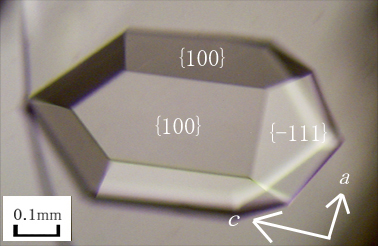
According to the reported crystallization condition [1], crystals of a hexagonal dipyramidal shape were obtained at 298 K and pH 5.5 (Figure 1). Indexes of crystal faces were determined to be {100} and {-111} based on precession photographs and facial angles. The macro-steps which may be attributed to bunching of the spiral steps were observed on {-111} by a differential interference microscope. Under high supersaturation, crystals of a squared shape surrounded by {-111} appeared at 298 K, and they tended to twin. On the other hand, crystals of parallelepiped plates appeared at 308 K.
Surface energies were estimated using the macrobond analysis [2] based on the PDB data of 1RNX. The surface energies of (-111), (100), and (001) increase in this order, which is consistent with the observed crystal shape.
[1]Alexander, A. et al. (1996). Biochemistry, 35, 15962-15972.
[2] Matsuura, Y. & Chernov, A. A. (2003). Acta Crystallogr., D59, 1347-1356.